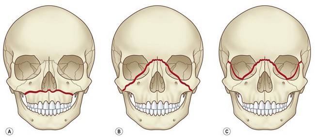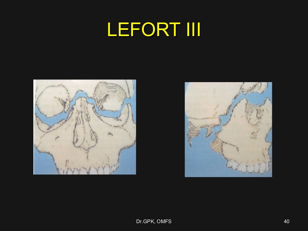

Type III fracture pattern extends posteriorly to the tuberosity or track approximate to the midline. The anterior limit of the fracture is between canine teeth which extend to the pyriform aperture. Type III is also called para-sagittal fracture which occurs in the thin part of the palate lateral to the attachment of vomer bone to the maxilla. Type III and IV fractures are the most common palatal fractures in adults. Type II palatal fracture is defined as sagittal fracture which is less common in adults. Anterior type I palatal fracture involves the incisor teeth and involving the posterior teeth it is defined as type 1b palatal fracture. Alveolar fracture is classified as type I palatal fracture in which it is categorized into two subcategories of anterior and posterolateral fractures. Computed tomographies (CTs) in coronal and axial views are helpful in detecting the palatal fractures. classified the palatal fracture into six patterns anatomically ( Figure 5). Bilateral zygomatic arches and zygomaticofrontal sutures and nasofrontal sutures should be fixed in severely displaced cases. The number of fixations is dependent on the extent of comminution and dislocation. Mobile mid-face and esthetic problems following Le Fort III fracture (dish-face deformity) are the main indications of ORIF treatment. Displaced Le Fort II fracture is treated by ORIF of bilateral infraorbital rims and zygomatic buttresses simultaneously using a miniplate to fix the nasofrontal suture. In the Le Fort I pattern lateral nasal walls and zygomatic buttresses are used to provide stability by four plates. The method of choice in the treatment of mobile maxilla with severe malocclusion is open reduction and internal fixation (ORIF). Closed technique could be performed by either maxillomandibular fixation (MMF) or skeletal suspension ( Figure 4). Minor maxillary displacement and malocclusion and low mobility of fractured segment are the indications of closed treatment. The decision to choose whether the open or closed technique in Le Fort fractures is dependent on the mobility of the maxilla and severity of maxillary displacement results in malocclusion. When the patient is stable, facial examination to detect the mid-face fractures is executed as follow. According to the possibility of spinal injuries in facial trauma patients stabilizing the cervical spine by a rigid collar is necessary until the spinal injury is ruled out.Īfter providing a secure airway, ATLS protocol can continued. The incidence rate of cervical spine trauma in pediatric facial fracture cases is almost 3.5% whilst this number is much higher in adult trauma patients. It is important to keep the airway open in mid-face fractures because there is always the potential of airway obstruction due to displacement of bones or severe bleeding in such cases.Ĭervical spine injuries are common in facial fractures. Intubation to secure the airway in instable mid-face fractures is the next step that should be considered in emergency patients. Packing should be used to control acute bleeding.

Removal of fractured teeth, clots, and loose dental crowns or dentures is important to open the oral airway. Hemorrhage and secretions may obstruct the oropharynx and nasopharynx. Airway obstruction should be evaluated as soon as possible since the mid-face is the beginning of the respiratory pathway. This chapter aims to present a comprehensive review of mid-face fractures types’ diagnosis and management.Īdvanced trauma life support (ATLS) is the first step that should be applied in emergency cases. Esthetic disfiguring trauma changes the whole mid-facial compartments.

Quality of life of the patients is influenced following unsuccessful management of mid-face fractures which lead to permanent functional problems. The treatment of mid-face fractures is complex due to the physiology and anatomy of mid-facial subunits. Diagnosis of the types of mid-face fractures is the first and basic step in management of mid-face trauma. Understanding the principles of mid-facial repair is the key to optimize the outcome.ĭiagnosing mid-face fractures is sometimes very difficult in emergency cases. The mid-face consists of vertical, horizontal, and sagittal pillars. The mid-face skeleton is important in providing a functional unit for respiratory, olfactory, vision, and digestive systems. The importance of mid-face is clear in function and esthetics. The other causes of facial fractures including mid-face trauma indicated in the literature are assaults, falls, sport injuries, and anima attacks. Motor vehicle accidents seem to be the first cause of mid-face fractures all around the word. Facial fractures are detected in almost 5–10% of trauma patients. Mid-face fractures are common in different populations.


 0 kommentar(er)
